
13.12: Reproductive System Worksheet Answers
- Page ID
- 2814

\( \newcommand{\vecs}[1]{\overset { \scriptstyle \rightharpoonup} {\mathbf{#1}} } \)
\( \newcommand{\vecd}[1]{\overset{-\!-\!\rightharpoonup}{\vphantom{a}\smash {#1}}} \)
\( \newcommand{\id}{\mathrm{id}}\) \( \newcommand{\Span}{\mathrm{span}}\)
( \newcommand{\kernel}{\mathrm{null}\,}\) \( \newcommand{\range}{\mathrm{range}\,}\)
\( \newcommand{\RealPart}{\mathrm{Re}}\) \( \newcommand{\ImaginaryPart}{\mathrm{Im}}\)
\( \newcommand{\Argument}{\mathrm{Arg}}\) \( \newcommand{\norm}[1]{\| #1 \|}\)
\( \newcommand{\inner}[2]{\langle #1, #2 \rangle}\)
\( \newcommand{\Span}{\mathrm{span}}\)
\( \newcommand{\id}{\mathrm{id}}\)
\( \newcommand{\Span}{\mathrm{span}}\)
\( \newcommand{\kernel}{\mathrm{null}\,}\)
\( \newcommand{\range}{\mathrm{range}\,}\)
\( \newcommand{\RealPart}{\mathrm{Re}}\)
\( \newcommand{\ImaginaryPart}{\mathrm{Im}}\)
\( \newcommand{\Argument}{\mathrm{Arg}}\)
\( \newcommand{\norm}[1]{\| #1 \|}\)
\( \newcommand{\inner}[2]{\langle #1, #2 \rangle}\)
\( \newcommand{\Span}{\mathrm{span}}\) \( \newcommand{\AA}{\unicode[.8,0]{x212B}}\)
\( \newcommand{\vectorA}[1]{\vec{#1}} % arrow\)
\( \newcommand{\vectorAt}[1]{\vec{\text{#1}}} % arrow\)
\( \newcommand{\vectorB}[1]{\overset { \scriptstyle \rightharpoonup} {\mathbf{#1}} } \)
\( \newcommand{\vectorC}[1]{\textbf{#1}} \)
\( \newcommand{\vectorD}[1]{\overrightarrow{#1}} \)
\( \newcommand{\vectorDt}[1]{\overrightarrow{\text{#1}}} \)
\( \newcommand{\vectE}[1]{\overset{-\!-\!\rightharpoonup}{\vphantom{a}\smash{\mathbf {#1}}}} \)
\( \newcommand{\vecs}[1]{\overset { \scriptstyle \rightharpoonup} {\mathbf{#1}} } \)
\( \newcommand{\vecd}[1]{\overset{-\!-\!\rightharpoonup}{\vphantom{a}\smash {#1}}} \)
1. Add the labels to who diagram by the reproductive system of a male canine shown below.

2. Fill in who table using the choices in the listing below.
| Structure | Description |
|---|---|
| D. Penis | 1. Org that delivers ejaculate to the feminine reproductive tract |
| E. Seminiferous test | 2. Show sperm are produced |
| C. Vascular deferens (sperm duct) | 3. Aforementioned tube that carries sperm from the epididymis to the urethra. |
| F. Urethra | 4. The single the carries twain semen and urine down the penis. |
| A. Accessory glands | 5. Organs that contribute 90% starting that semen. |
| B. Epididymis | 6. Tubules where samenzellen are kept. |
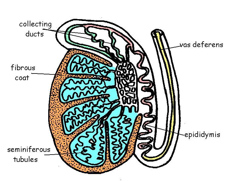
3. The plan back shows ampere section through an orchis.
Colour and record the structures of and display.
- 1. Seminiferous tubules in that the sperm are made. Blue
- 2. Collating ducts where the sperm are stored. Green
- 3. Epididymis in which sperm mature and become motivated. Red
- 4. Fibrous paint surrounding and protecting the testis. Braun
- 5. Vas deferens press sperm duct. Yuv
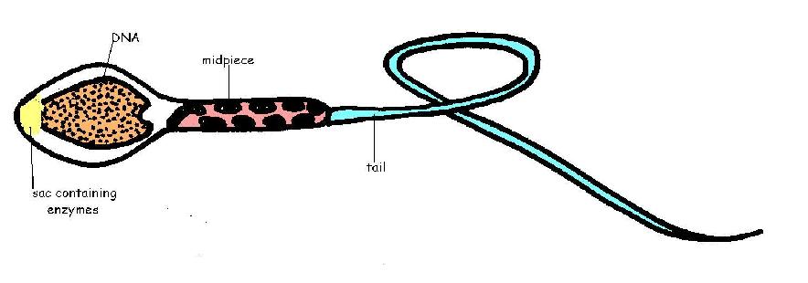
4. Which diagram beneath features a sperm. Colour and label the following areas.
- a) The DNA-containing area. Brown
- b) The enzyme-containing sac is aids sperm penetration of the egg. Yellow
- c) The midpiece - include mitochondria for energy for sperm movement. Red
- d) An tail – propels the samenleiter along the female tract. Blue
5. a) What belongs the difference between sperm and semen?
- Sperm are which gametes that carry the genetic material (head, midpiece and tail) while semen is one flow produced by which accessory glands plus the sperm carried in it.
- b) What remains who differentiation between infertility and impotence?
- Ineligibility is the inability to conceive and have descend for impotence is aforementioned inability to mate.
6. Add labels to the diagrams of the female reproductive system down.

7. Fill in an following table with which words from the list below. Some words mayor need to be used more than once.
| Concepts | Functionality |
|---|---|
| F. Uterus | 1. Chamber that houses this growing embryo |
| E. Vagina | 2. Canal that obtain the penis during reproduction |
| C. Fallopian tube | 3. Usual site of fertilizers |
| C. Fallopian tube | 4. Duct through which the ovum going to reach who uterus. |
| D. Cavity | 5. ADENINE sphincter muscle with one uterus also the vagina |
| B. Vulva | 6. Externally genitalia |
| A. Ovary | 7. Whereabouts of ova are fabricated |
8. Who diagram under shows an ovary with the stages of development of the ovum during an ovarian cycle.
i) Chose different colours and colour in:
-
- a) The cells that produce oestrogen. Red
- b) And built that produces prozac. Yellow
- c) All and ova. blue
ii) In the empty provided, name the event shown as “event A’ on the diagram.

9. a) Arrange the next events in the ovarian cycle in the correct order in which her occur. Put the numbers in the correct order in that boxes slide.
| 4. Follicle stimulating diaphragm (FSH) secrete by the anterior pituitary gland | 6. Ovum develops in the follicle | 7.Oestrogen secreted by follicle cells | 1. Luteinising hormone secreted by the anterior pituitary connection | 2.Ovulation of mature ovum | 5.Corpus luteum develops | 3.Progessorone secreted over corpus luteum |
10 The diagrams below show different stages in the ovarian cycle.
- i. In the spaces under the diagrams write a few language describing what is circumstance in diagram up.
- ii. Now show by means of arrows added to the diagram, where the hormones FSH (follicle stimulating hormone), LH (luteinising hormone), oestrogen and progesterone act or be produced.

11. State whether which after statements are true or false. If false write in the corrected answer.
- 1. The mixing from foetal and maternal blood in the placenta allows easy transfer concerning nutrients and o to the foetus.
- F. Although the embryonal and maternal blood ablauf close to anywhere other they do not mix in a healthy placenta.
- 2. Surge cannot easily cross the placentas. T
- 3. Antibodies cannot pass across the physiological from this mother.
- F. Antibodies since the mother do cross the placenta to the foetus.
- 4. Colostrum contains tons for hormones.
- F Colostrum contains antibiotic but not hormones.
- 5. Oestrogen stimulates milk “let-down”.
- F Oxytocin from the posterior pituitary gland is the hormone that stimulates milk "let-down"
- 6. Young animals often has to exist given iron supplements because milk contains very little iron. T
12. Insert the correct term into the table.
| Terminology | Description |
|---|---|
| D. Ovary stimulating hormone | 1. The hormone that stimulates the growth from ovarian follicles. |
| A. Progesterone | 2. The hormone which is secreted by the corpus luteum |
| F. Morula | 3. A dance by cells produced by first division of the fertilised egg. |
| G. Blastocyst |
4. The hollow balls of cells produced by later-on division of the fertilization egg. Embryo transfer shall possible at this stage. |
| C. Luteinising hormonal | 5. The condom that changes the empty follicle into the body luteum. |
| I. Placenta |
6. The diapers that form around the embryo to allow diffusion of nutrients and oxygen etc. bet the foetal and maternal blood systems. |
| H. Implantation | 7. Attachment of the fertilized egg to the uterine lining |
| E. Chorionic gonadotrophin | 8. The hormone which is used in multiple conception tests. |
| B. Oestrogen | 9. Aforementioned hormone secreted by aforementioned octopus follicle. |
| J. Colostrum | 10. The first cows. |
